Atlas of Human Anatomy uses Frank H. Netter, MD's detailed illustrations to demystify this often intimidating subject, providing a coherent, lasting visual vocabulary for understanding anatomy and how it applies to medicine. This 5th Edition features a stronger clinical focus-with new diagnostic imaging examples-making it easier to correlate anatomy with practice. Student Consult online access includes supplementary learning resources, from additional illustrations to an anatomy dissection guide and more. Netter. It's how you know.
Frank H. Netter Boeken
25 april 1906 – 17 september 1991
Frank H. Netter was een arts en vooraanstaand medisch illustrator die de werelden van kunst en geneeskunde verbond. Zijn gedetailleerde en nauwkeurige anatomische tekeningen hebben een diepgaande invloed gehad op medisch onderwijs en praktijk. Hij gebruikte zijn artistieke talent om visueel boeiende werken te creëren die talloze studenten en beoefenaars hielpen de complexiteit van het menselijk lichaam te begrijpen. Zijn blijvende nalatenschap vormt een hoeksteen van medische illustratie en duidelijkheid in anatomisch begrip.


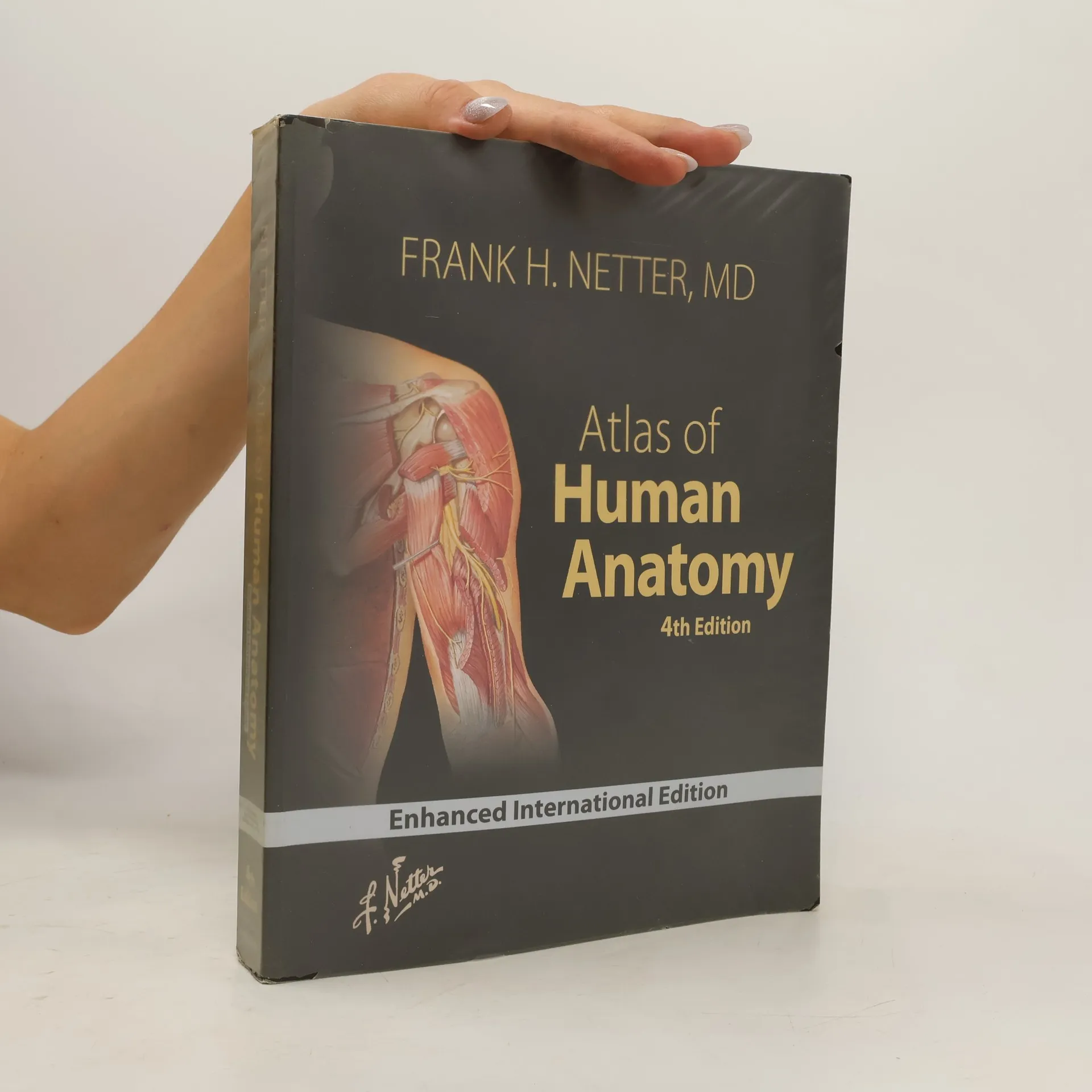
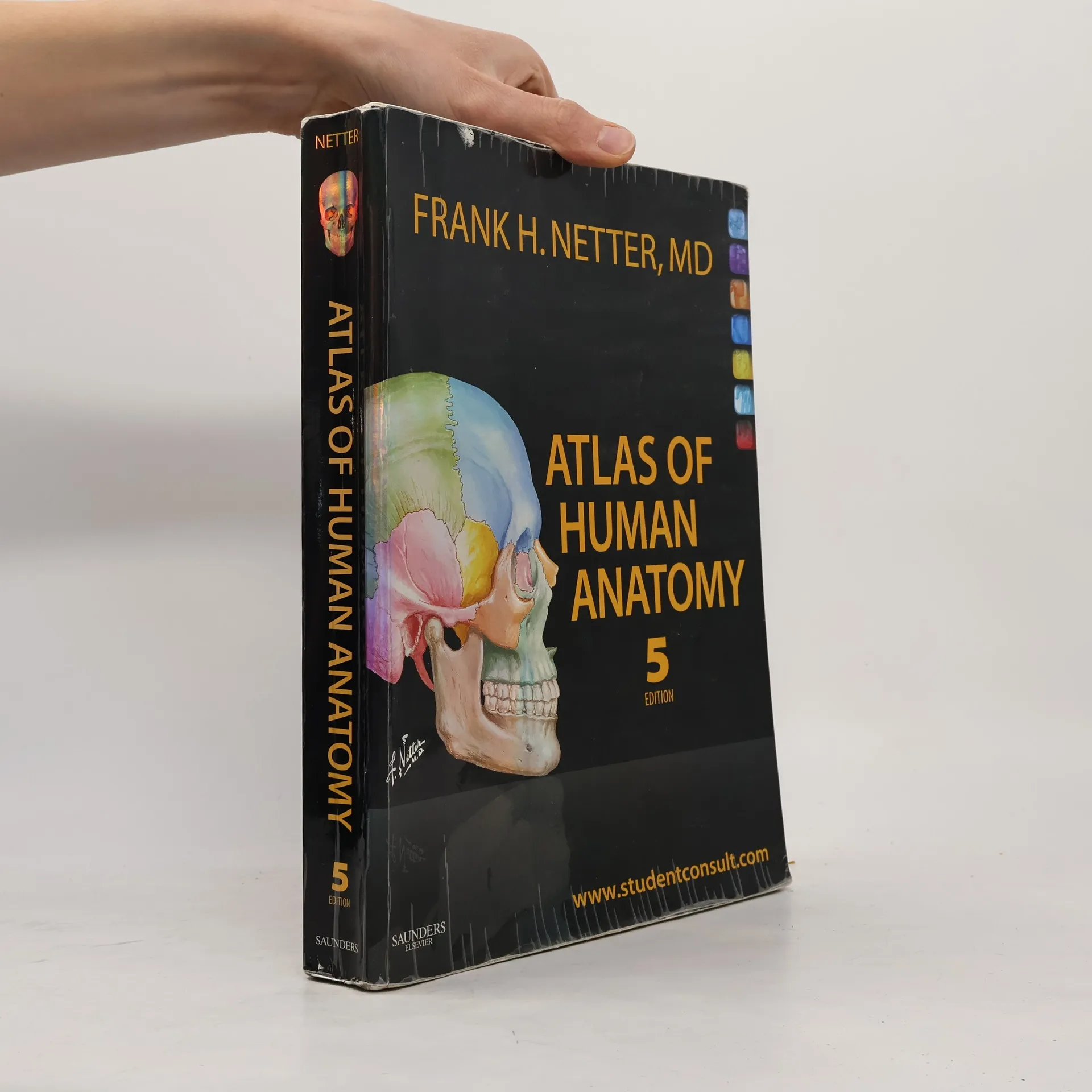
Atlas of Human Anatomy
- 662bladzijden
- 24 uur lezen
Accompanying CD-ROM entitled: Interactive atlas of human anatomy.
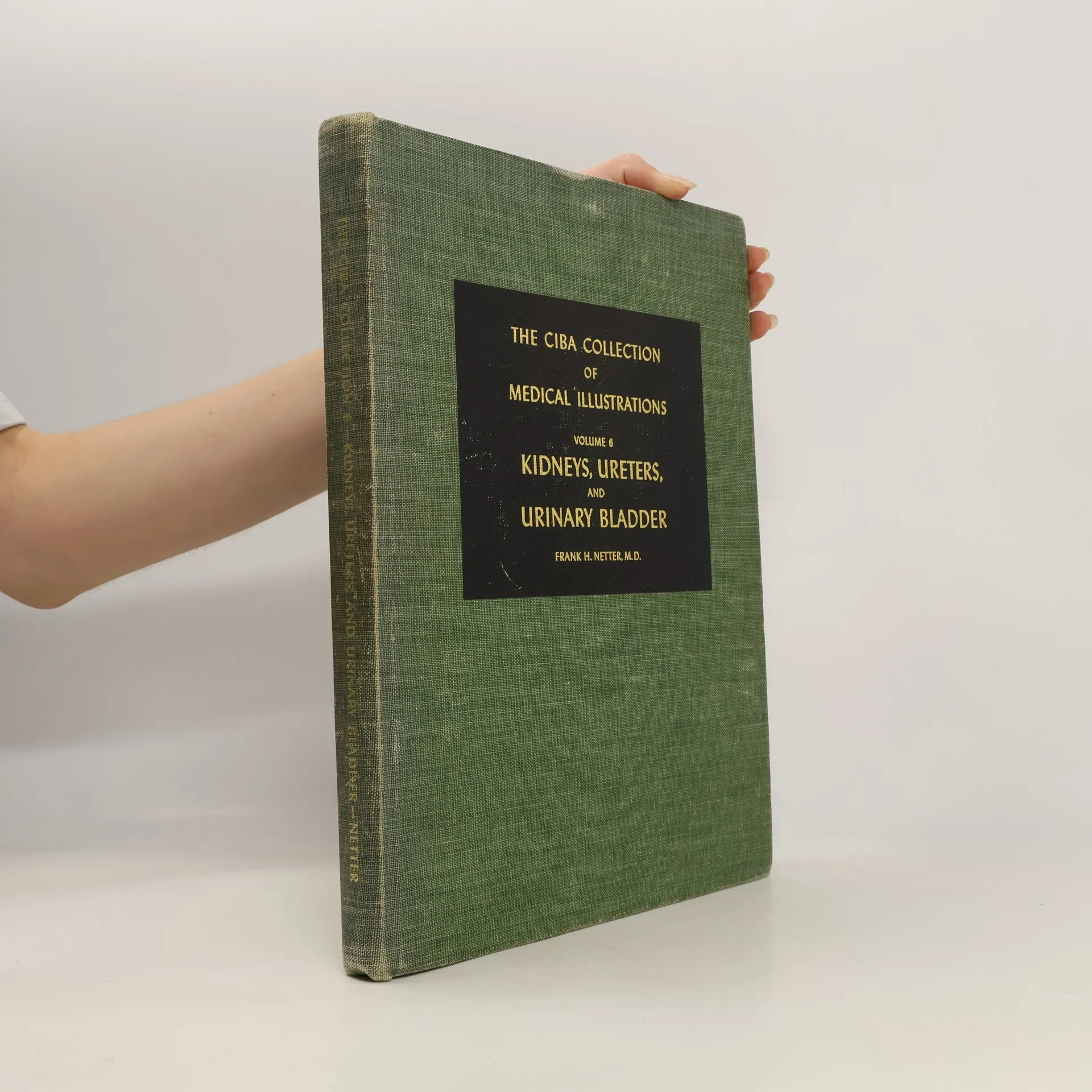
The most critically acclaimed of all of Dr. Frank H. Netter's works, this fully illustrated single book from the 8-volume/13-book reference collection includes: hundreds of world-renowned illustrations by Frank H. Netter, MD; informative text by recognized medical experts; anatomy, physiology, and pathology; and diagnostic and surgical procedures.
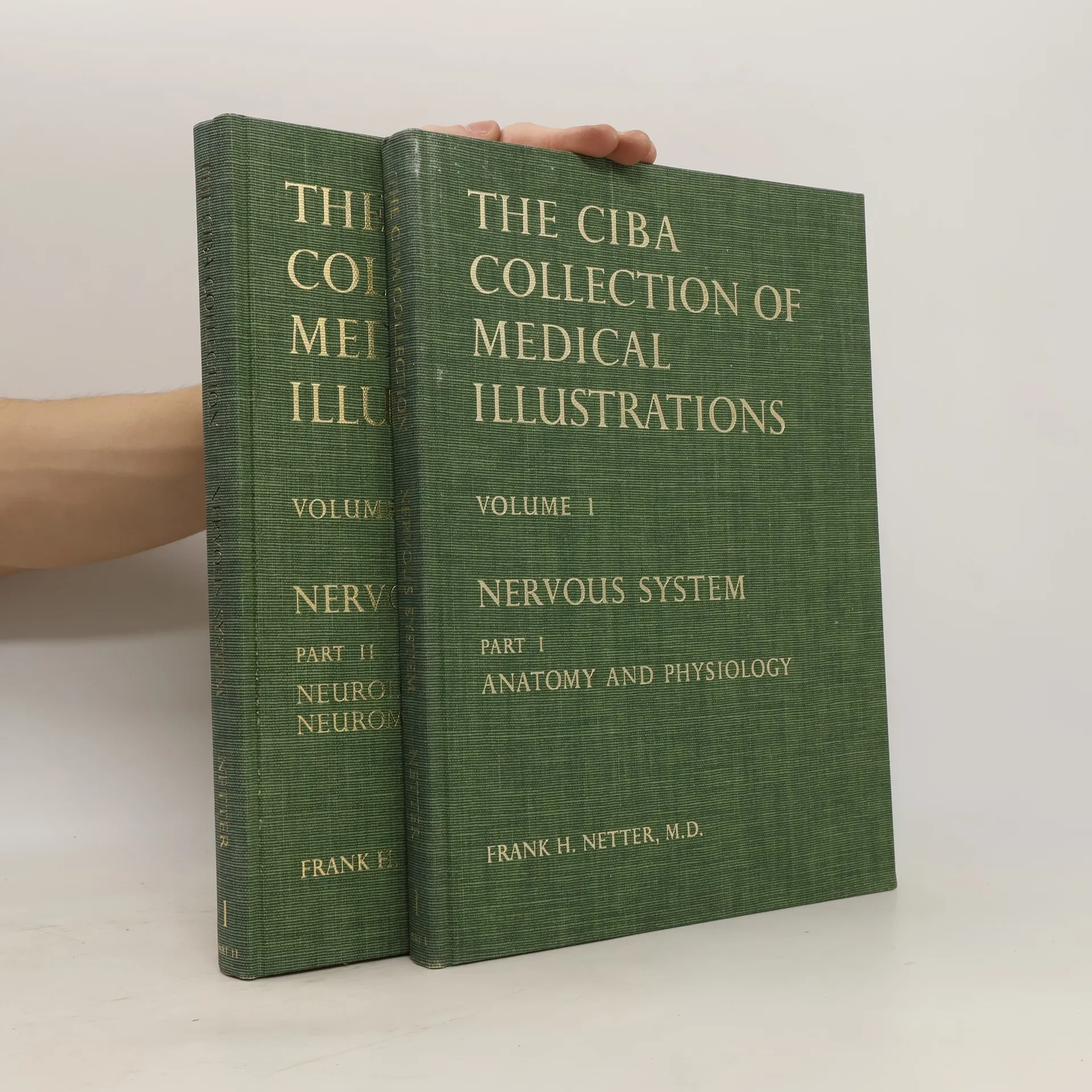
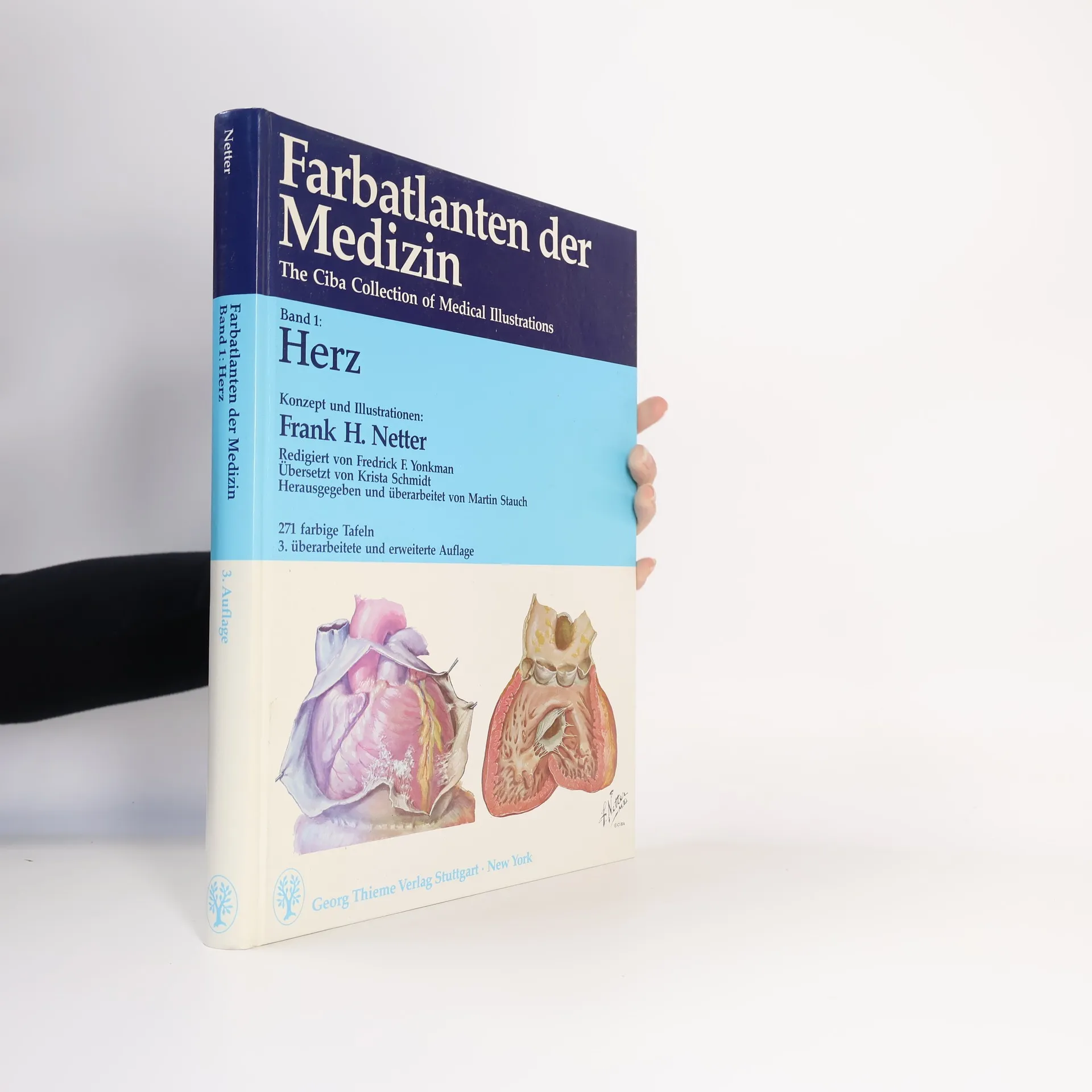
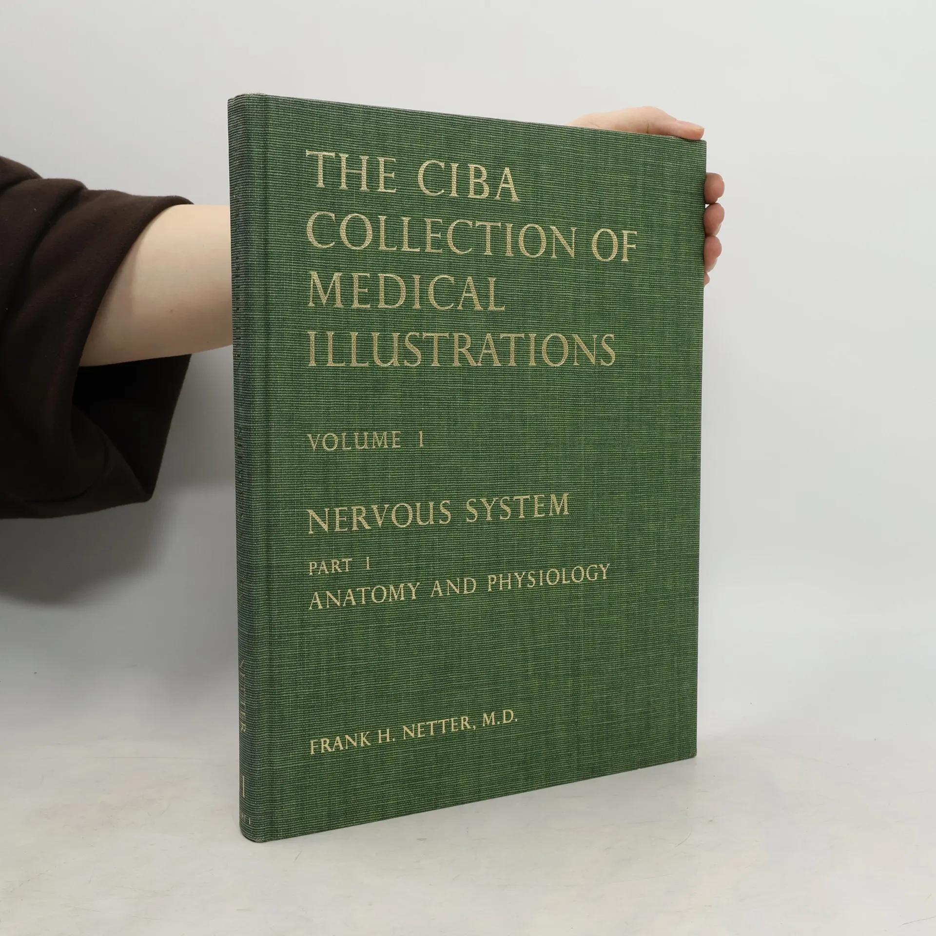
The CIBA Collection of Medical Illustrations
Volume 1. Nervous System. Part I. Anatomy and Physiology
Iconic medical illustrator Netter MD shows his skills best in this volume on the nervous system. This volume is one of a master set of thirteen books.
Focusing on the needs of students and clinical professionals, this anatomy atlas offers clear, detailed illustrations of the human body, organized by region. Its unique approach highlights key anatomic relationships relevant to clinical practice, featuring over 550 meticulously crafted plates alongside selected radiologic images for common perspectives. Created by clinicians for clinicians, it serves as an essential resource for those learning anatomy, participating in dissection labs, or refreshing their knowledge.
The Ciba collection of medical illustrations 1. Part II.
- 256bladzijden
- 9 uur lezen
With hundreds of illustrations, this title features informative text by medical experts; anatomy, physiology, and pathology; and diagnostic and surgical procedures.
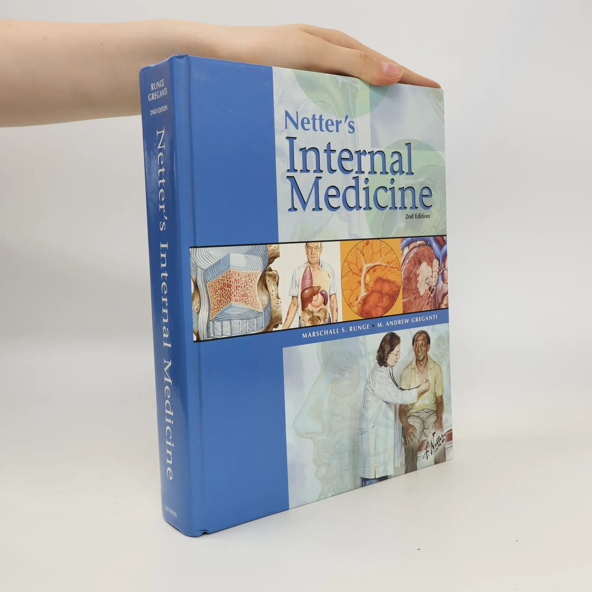
Gain fast, easy visual access to the problems most often encountered in practice! This resource combines hundreds of exquisite Netter images - including several new paintings created especially for this book - with concise summaries of the most current medical thinking on common diseases/conditions, diagnostics, treatments, and protocols - for a single easy-to-use quick-reference guide. Instructive and memorable Netter plates provide a rich visual understanding of every concept. The result is a superb source for ongoing clinical reference as well as patient and staff education. Offers quick access to expert medical thinking on common diseases/conditions, diagnostics, treatments, and protocols. Presents more than 500 exquisite illustrated plates by master illustrator Frank H. Netter and other artists working in the Netter tradition to enhance your understanding of the material. Presents nearly 40 new chapters, many expanded chapters, and several new images to reflect the state of internal medicine today-including increasingly common issues like bariatric surgery and posttraumatic stress syndrome. Offers more tables and algorithms for enhanced at-a-glance" guidance. Features annotated citations for additional resources, including websites and other key sources for practice guidelines and patient education and support. Presents annotated evidence from key studies that have shaped the current standard of care.
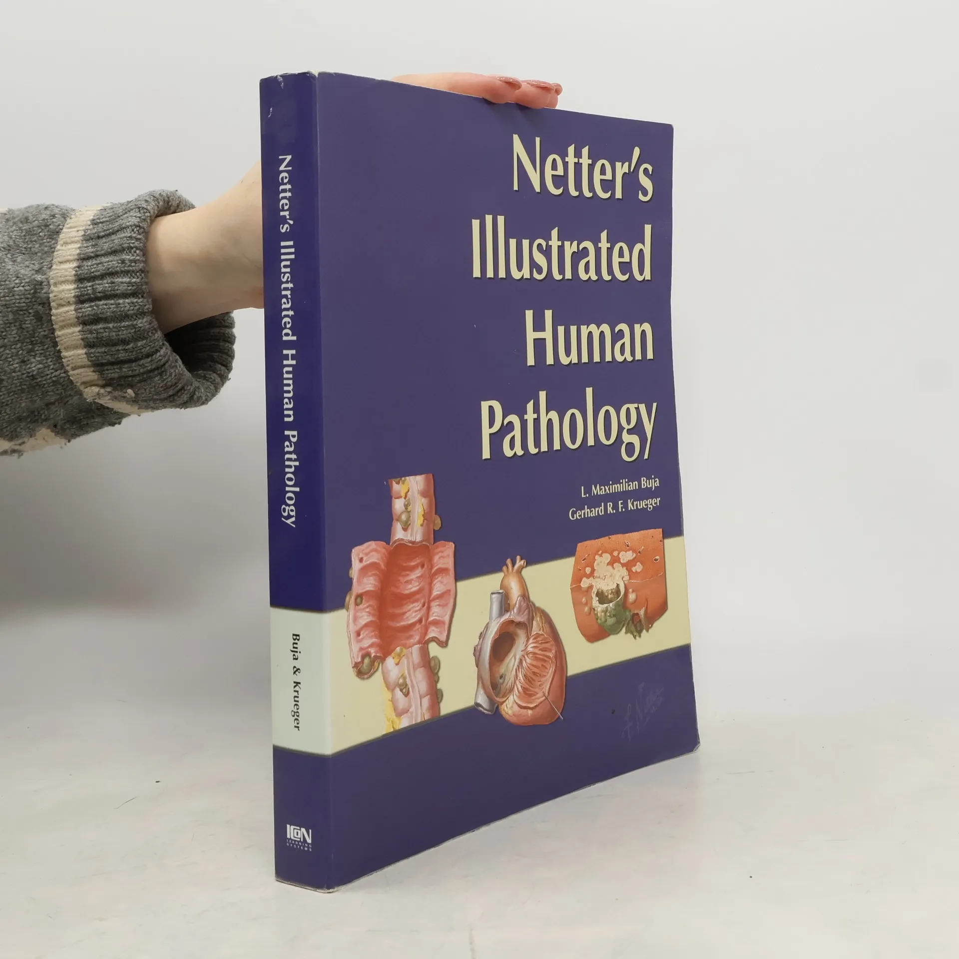
Netter's Illustrated Human Pathology
- 527bladzijden
- 19 uur lezen
Netter's Atlas of Human Pathology is a visually vibrant approach to help students gain critical insight into the structure-function relationships and the pathological basis of human disease. It provides clear and succinct representations of common human diseases by relating anatomical changes to the functional and clinical manifestations of disease and their underlying causes and mechanisms. Features more than 380 classic Netter and new Netter-sytle images, gross and microscopic photographs and tables.The book consists of 12 organ-specific chapters - each containing precisely rendered illustrations of pathological processes and diseases accompanied by relevant text to expand knowledge and clarify understanding. Comparative data about similar disease processes are summarized in tables.Netter's Illustrated Human Pathology The Atlas offers a superb complement to more comprehensive textbooks and presentations of pathology, including course syllabi. It can also be used as an adjunct for study of gross and microscopic pathology specimens in laboratory exercises, and makes a great review resource for students, medical residents, physicians and other healthcare professionals.
Netter Atlas of Human Anatomy: Classic Regional Approach (hardcover)
Professional Edition with NetterReference Downloadable Image Bank
- 712bladzijden
- 25 uur lezen
This atlas serves as a comprehensive resource for clinical professionals and students, offering detailed illustrations of human anatomy from a clinician's perspective. It features over 550 plates and radiologic images, highlighting key anatomical relationships essential for training and practice. New illustrations and nerve tables enhance coverage of clinically significant areas. The content is guided by expert anatomists and adheres to international anatomical standards, making it a vital tool for dissection labs and patient education. Additionally, it provides access to a downloadable image bank for personal use.
Netter Atlas of Human Anatomy: A Systems Approach
- 712bladzijden
- 25 uur lezen
The Netter Atlas of Human Anatomy is an essential resource for students and clinical professionals. It offers detailed illustrations of the human body, emphasizing important anatomical relationships for clinicians. With over 550 exquisite plates and selected radiologic images, it serves as a comprehensive guide for learning and refreshing anatomy knowledge.
Netter's Head and Neck Anatomy for Dentistry
- 659bladzijden
- 24 uur lezen
Netter's Head and Neck Anatomy for Dentistry, by Neil S. Norton, PhD, uses more than 600 full-color images from the Netter Collection to richly depict all of the key anatomy that's relevant to clinical practice. This new edition takes your knowledge further than ever with more Netter illustrations; addition of over 20 cone beam CT images; new chapters on the upper limbs, thorax, and abdomen; and more than 100 multiple-choice questions. Whether for your dental anatomy course, board review, or as a handy reference in your dental office, this concise, visual guide is an excellent anatomy atlas and quick reference for students and professionals in dentistry and dental hygiene. Identify clinically relevant anatomy with Netter illustrations highlighted and modified for dentistry. See the practical important of anatomy from illustrated clinical examples in each chapter. Review essential concepts easily with tables that display the maximum amount of information in an at-a-glance format. Master anatomy for the head and neck and beyond, including upper limbs, thorax, and abdomen. Stay current on hot topics like cone beam CT imaging, intraoral injections, and anesthesia. Recognize the context and clinical relevance of head and neck anatomy through additional coverage of dental procedures. Prepare effectively for the dental boards with over 100 multiple-choice questions.
Netter's Atlas of Human Embryology
- 267bladzijden
- 10 uur lezen
Here's a rich pictorial review of normal and abnormal human prenatal development. For each body system or region, you'll find a brief description of the developmental plan, with key concepts and terminology, followed by discussions of histological principles, the classification of congenital defects, and basic cellular, molecular, and genetic concepts.An emphasis on morphological patterns in the embryo and fetus makes it easy to understand the structure and function of the adult body and the embryonic basis of birth defects.Summary tables and terminology sections at the end of each chapter, plus an appendix with all major congenital defects and their embryonic basis, make it easy to review course material and prepare for the USMLE.
The most critically acclaimed of all of Dr. Frank H. Netter's works, this 8-volume/13-book reference collection includes: hundreds of world-renowned illustrations by Frank H. Netter, MD; informative text by recognized medical experts; anatomy, physiology, and pathology; and diagnostic and surgical procedures.
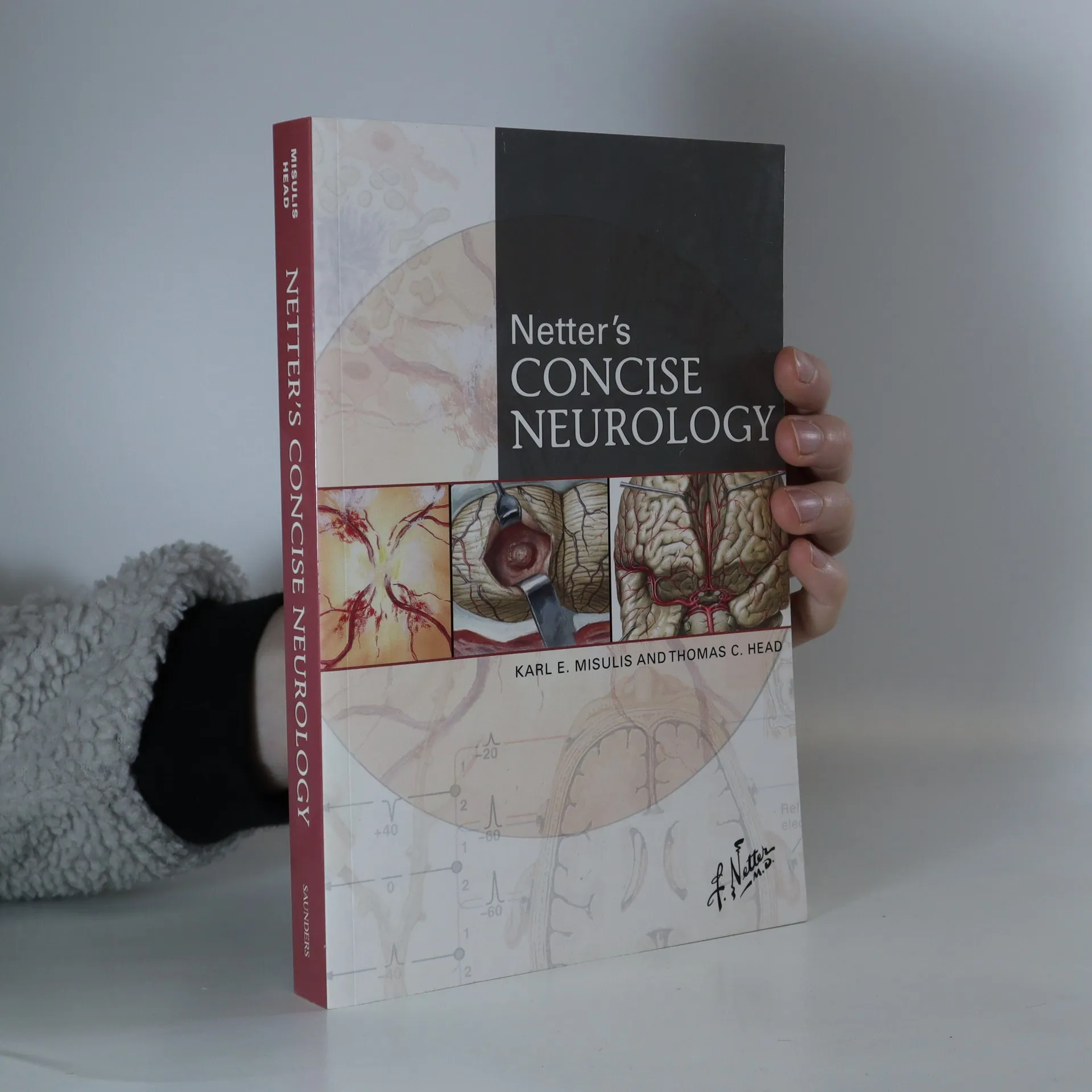
Netter's concise neurology
- 586bladzijden
- 21 uur lezen
More than 200 exquisite, hand-painted illustrations - created by, and in the style of, master medical illustrator Frank H. Netter, MD - capture the essential clinical aspects of over 200 major neurologic disorders seen in hospital and office practice. A masterful combination of artwork, succinct text, and tables, together with a highly compact format, deliver quick and convenient access to vital clinical knowledge!
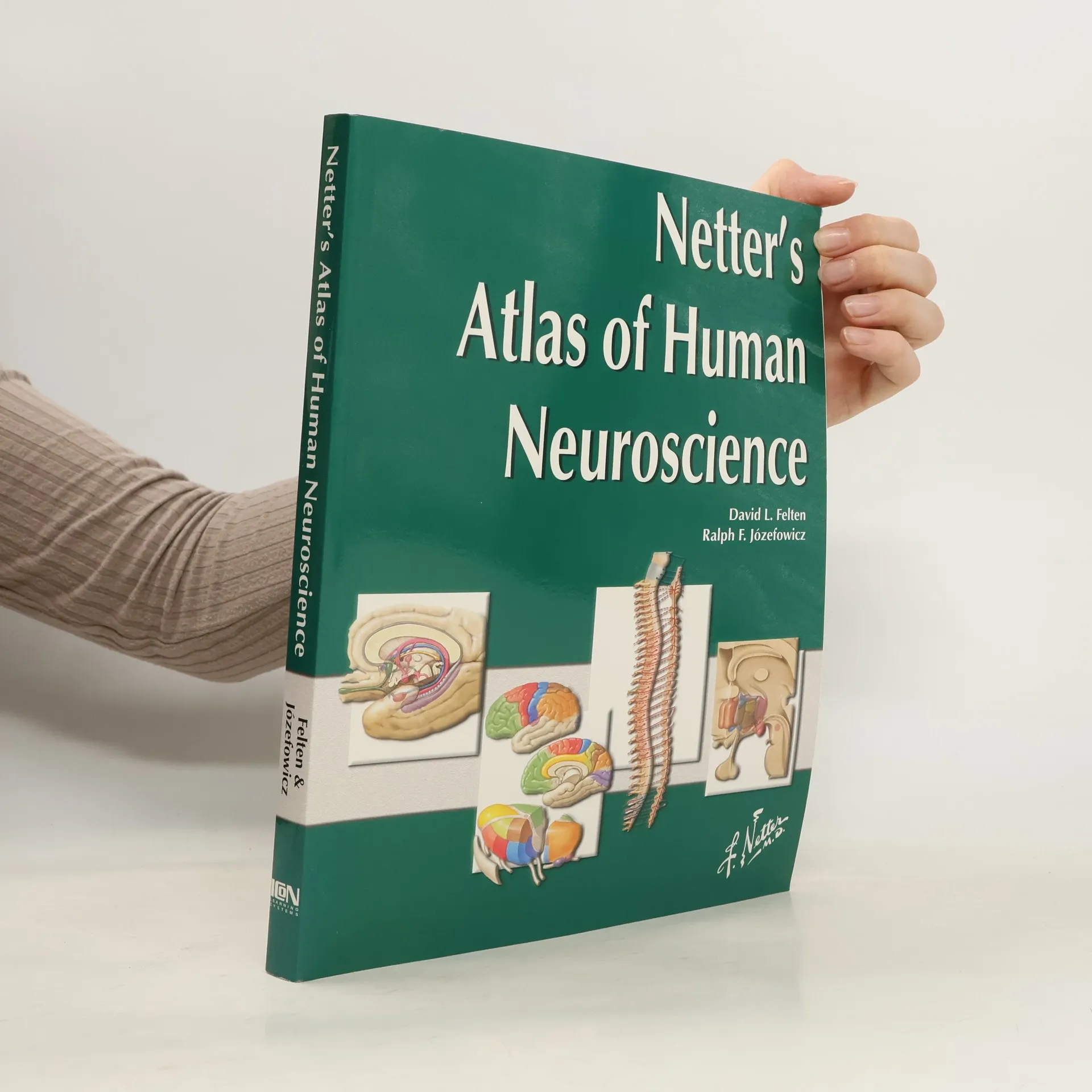
Netter Basic Science: Netter's Atlas of Human Neuroscience
- 310bladzijden
- 11 uur lezen
This atlas combines the precision and beauty of 325 Netter and Netter-style illustrations with updated information to reflect our growing understanding of the many regions and systems of the brain, spinal cord, and periphery. Concise neuroscience atlas using Netter illustrations to highlight key neuroanatomical concepts and clinical correlations. The single best source of illustrations of the nervous system, with comprehensive up-to-date information in a succinct and useful format, reflecting current understanding of the nervous system. Format of color plate with legend -- legends included on the same page as the illustrations to prevent the need for turning pages back and forth. Several tightly organized tables included to eliminate the need for long or detailed figure descriptions or text. These tables are useful aides to student learning. Schematic cross-sectional brain stem anatomy, and side-by-side comparisons of horizonal sections, CTs and MRs, eliminate the need for an additional purchase of a detailed neuroanatomy atlas. Netter's well-recognized and aesthetically pleasing neurosciences illustrations updated to reflect today's science.
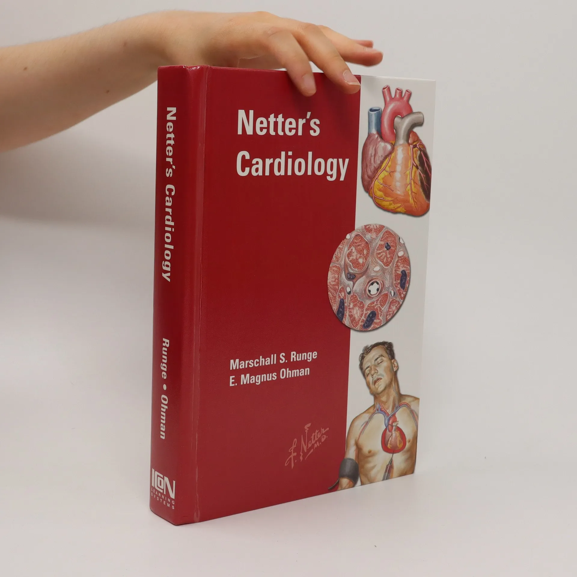
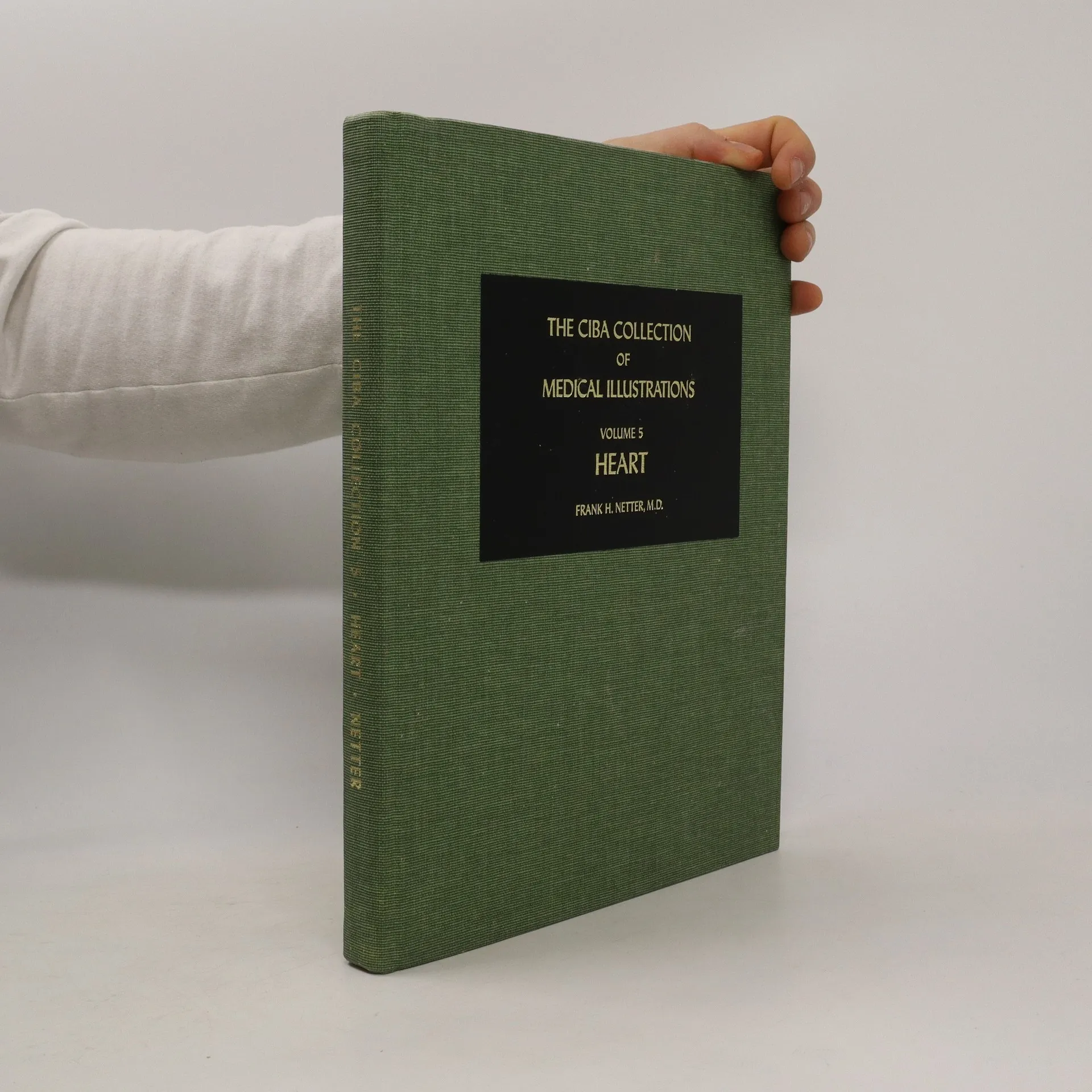
Netter's cardiology
- 664bladzijden
- 24 uur lezen
Netter's Cardiology offers residents and clinicians an easily accessible and practical resource on all aspects of the field. This text presents the essentials of clinical practice in cardiovascular disease merged with the incomparible illustrations of Frank H Netter.
The Netter Collection of Medical Illustrations Complete Package
- 9bladzijden
- 1 uur lezen
The collection offers an in-depth visual exploration of human anatomy and clinical pathology across nine updated volumes, featuring Dr. Frank H. Netter's renowned illustrations alongside new artwork. Each volume integrates core concepts of anatomy and physiology with clinical applications, making complex information accessible for students and professionals in health and medicine. The inclusion of an eBook enhances the learning experience, allowing for interactive features like searching and note-taking, solidifying this series as an essential educational resource.
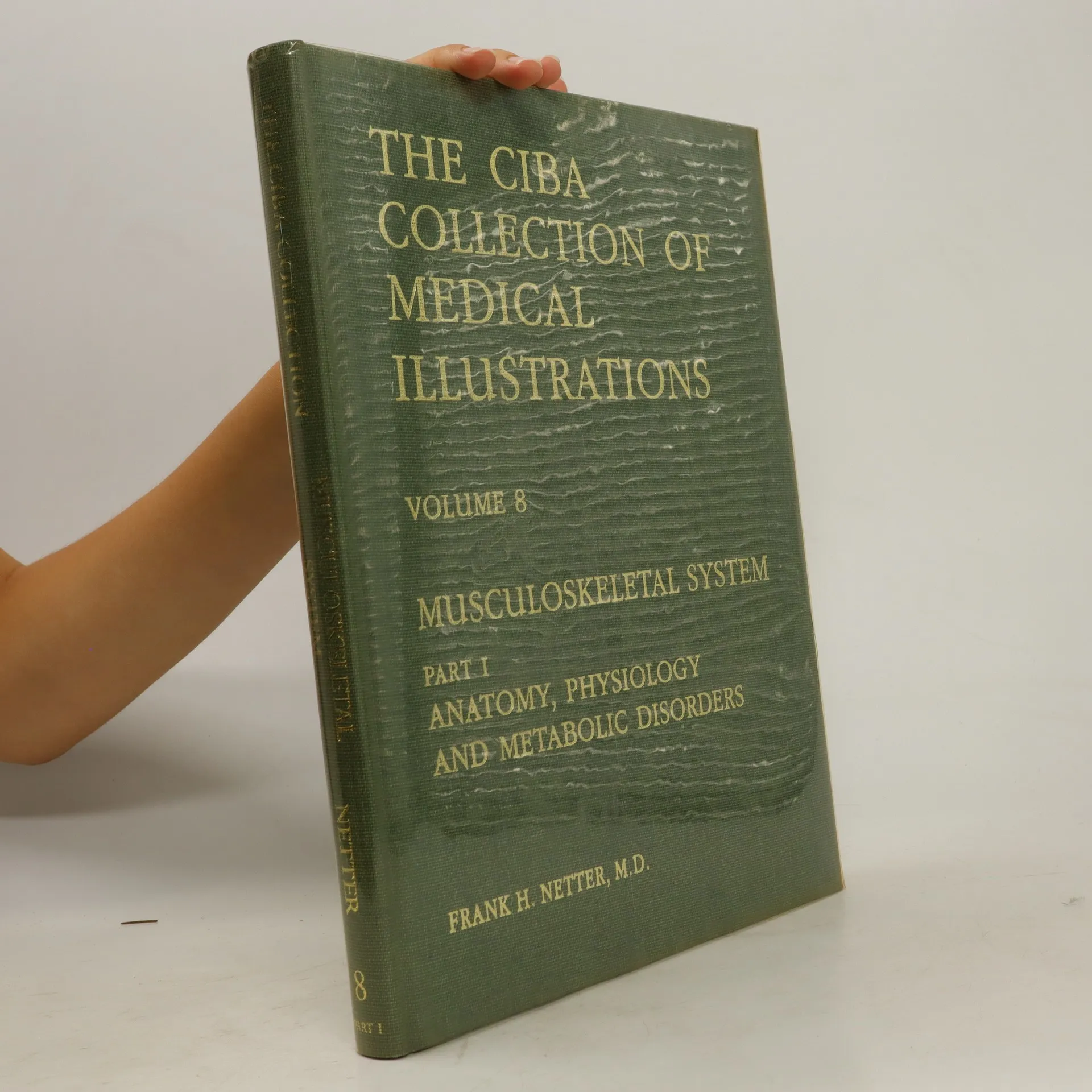
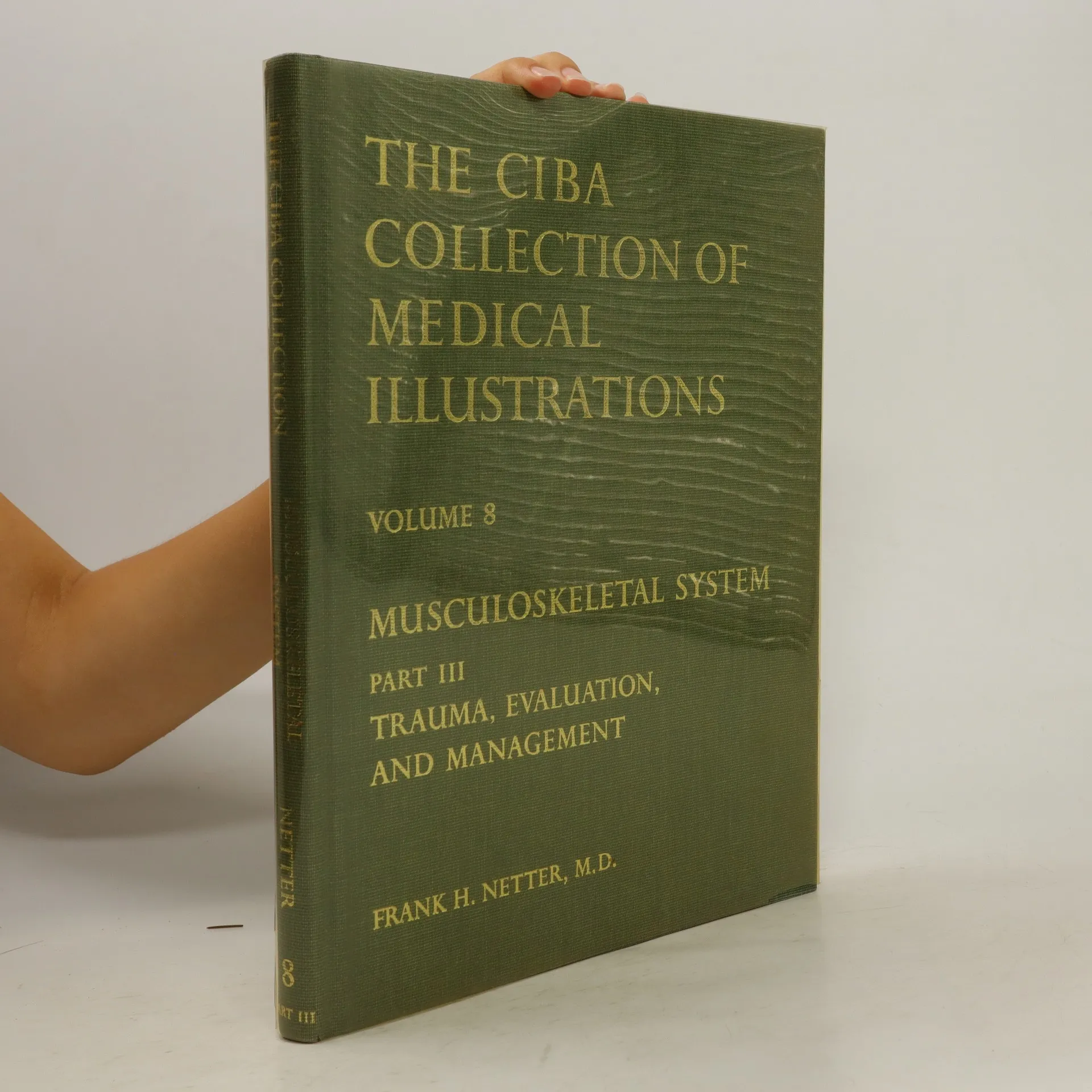
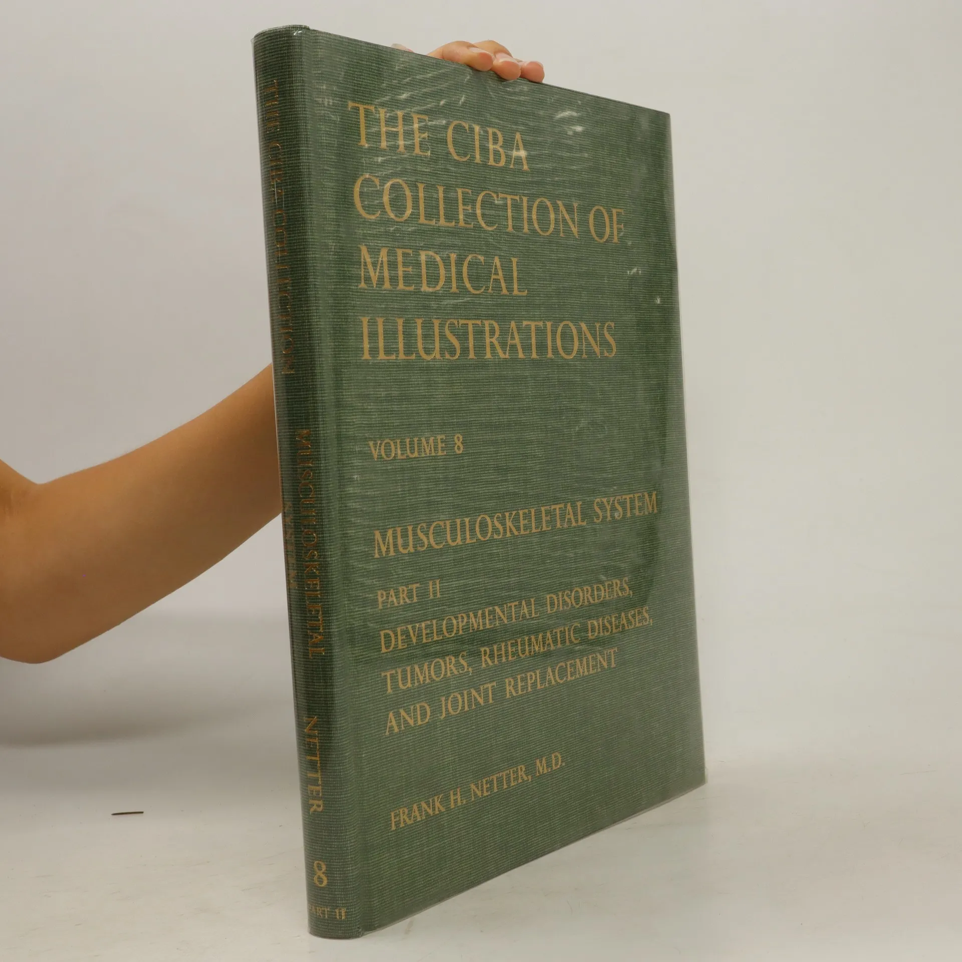
The Ciba Collection of Medical Illustrations 8
- 22bladzijden
- 1 uur lezen
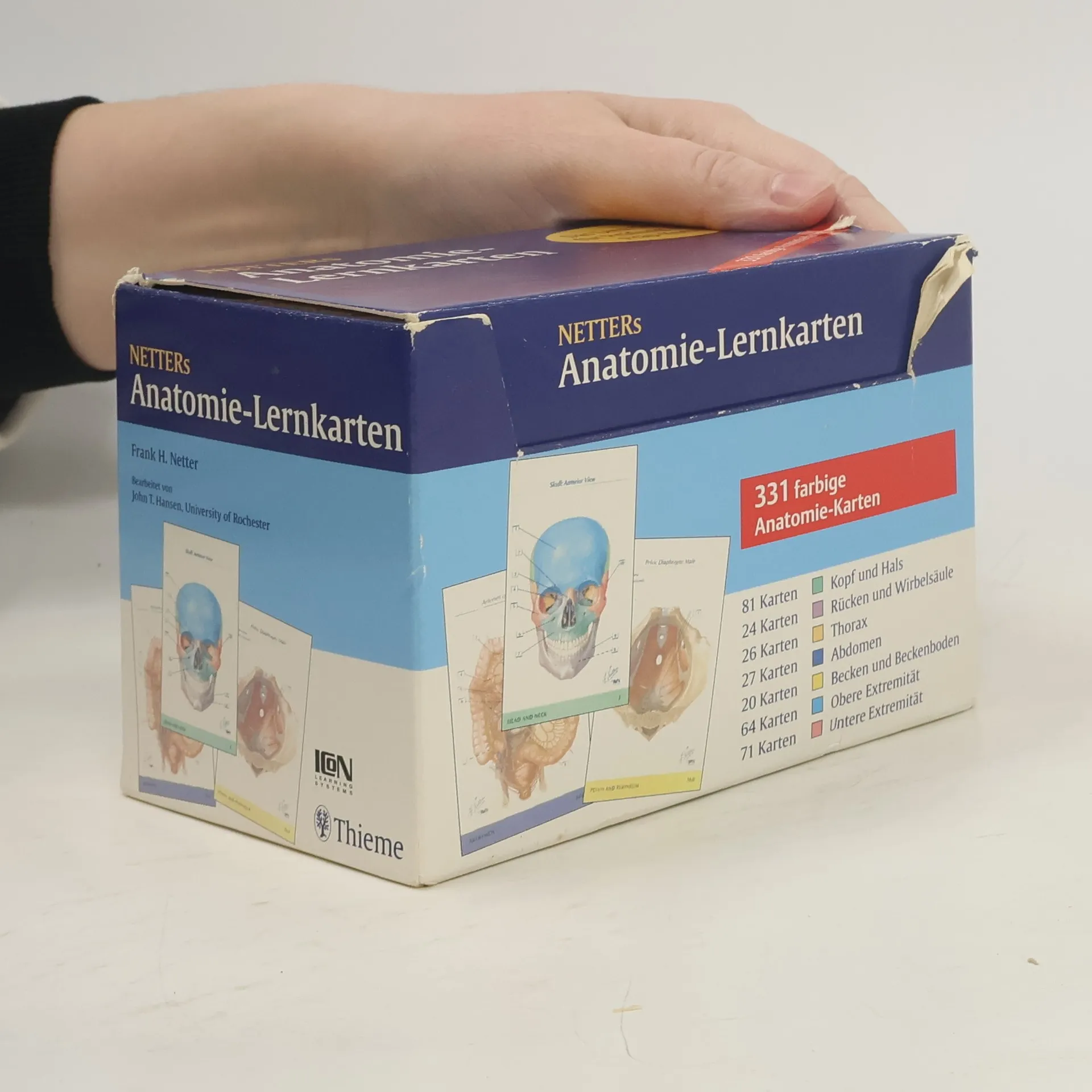
Netter's Anatomy Flashcards in englischer Sprache sind eine einzigartige Ergänzung zum Atlas und erleichtern das Lernen - überall und jederzeit. Beschriftungen der Zeichnungen lateinische Nomenklatur ansonsten alle Texte englisch! Anatomie erfolgreich lernen mit 324 perfekt illustrierten farbigen Lernkarten. Geordnet nach Körperregionen: Kopf und Hals, Rücken und Wirbelsäule, Thorax, Abdomen, Becken und Dammregion, Obere Extremität, Untere Extremität. Auf der Vorderseite jeder Karte findet sich eine detaillierte anatomische Zeichnung, auf der Rückseite die Bezeichnung der Strukturen gemäß Lateinischer Nomenklatur!
This book is the Ciba collection of medical illustrations and musculoskeletal system with developmental disorders, tumors, rheumatic diseases and joint replacement information.
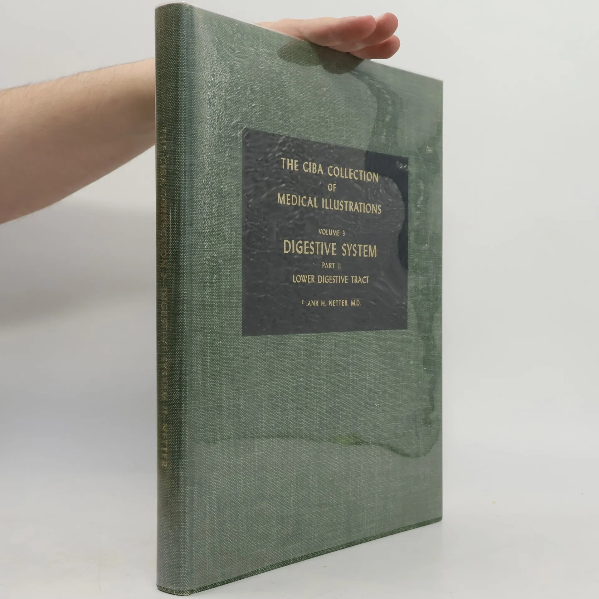
Digestive System 3
Part II Lower Digestive Tract
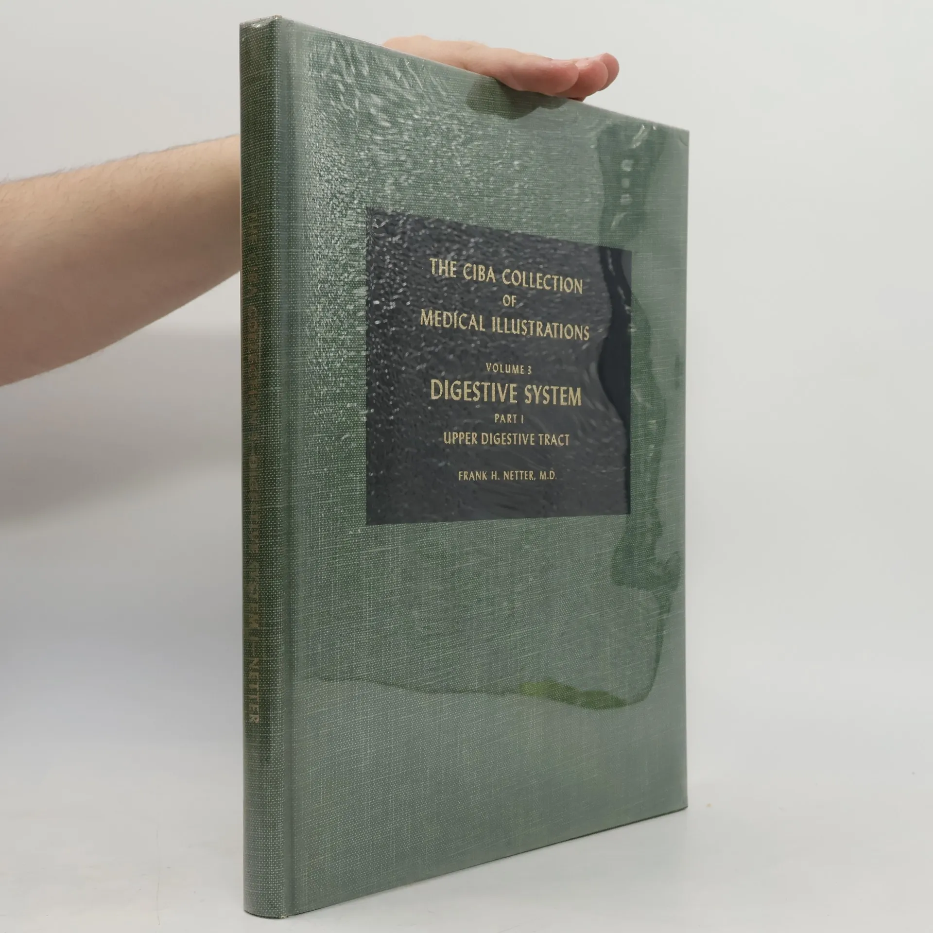
Digestive System 3
Part I: Upper Digestive Tract
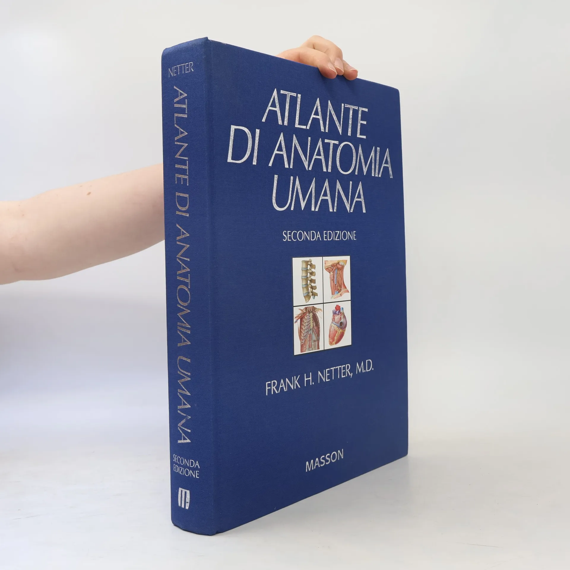
Atlante di anatomia umana. Con CD-ROM - Seconda edizione
- 574bladzijden
- 21 uur lezen
The Ciba Collection of Medical Illustrations 3
Digestive System III
The most critically acclaimed of all of Dr. Frank H. Netter's works, this fully illustrated single book from the 8-volume/13-book reference collection hundreds of world-renowned illustrations by Frank H. Netter, MD; informative text by recognized medical experts; anatomy, physiology, and pathology; and diagnostic and surgical procedures.
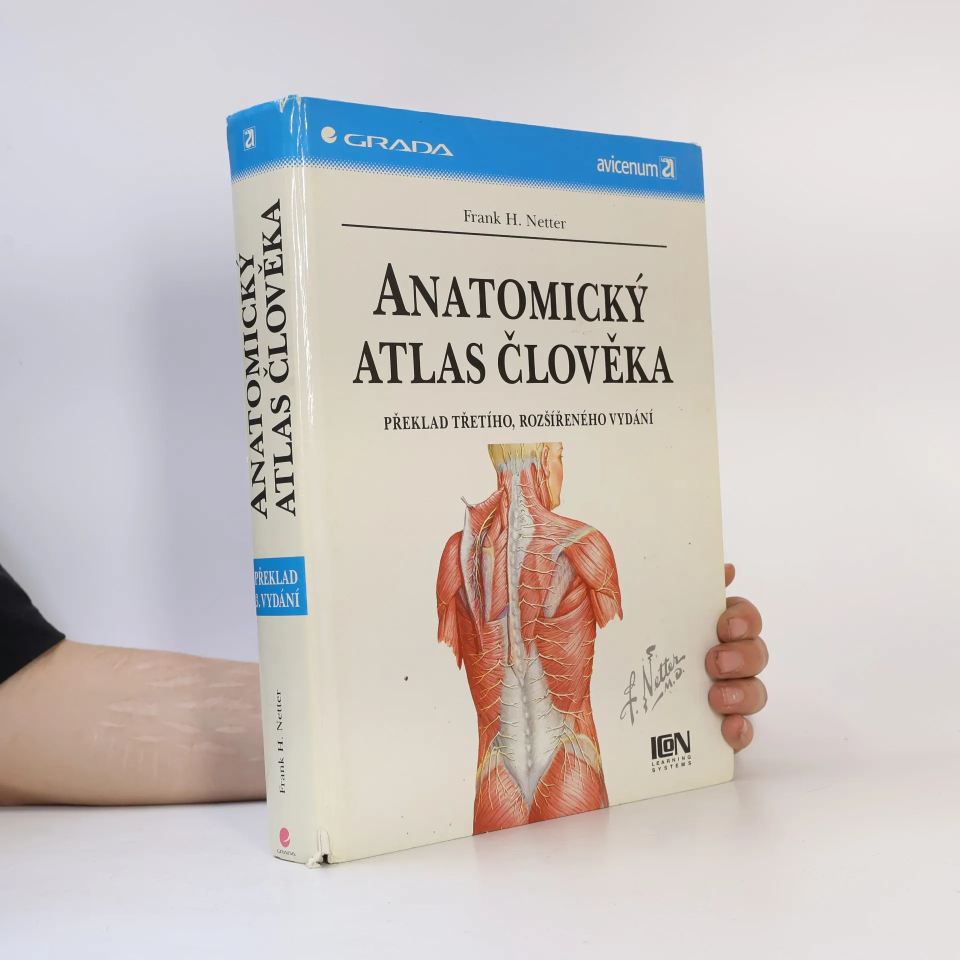
Známý ilustrovaný anatomický atlas, který je oblíben jak u studentů medicíny, tak u jejich vyučujících. Učebnice obsahuje 531 barevných anatomických tabulí, každá kapitola je doplněna tabulkami svalů. Vynikající a názorné ilustrace posunují tuto knihu od učebnice anatomie k uměleckému dílu.
Anatomický atlas člověka
- 628bladzijden
- 22 uur lezen
Světově uznávané dílo - vrchol anatomické vědecké ilustrace, přeložen do 16 jazyků. Anatomii vyobrazuje jasně, realisticky a vyváženě. Více než 540 barevných velmi názorných ilustrací, radiografy, snímky výpočetní tomografie, CT angiogramy, snímky magnet. rezonance.
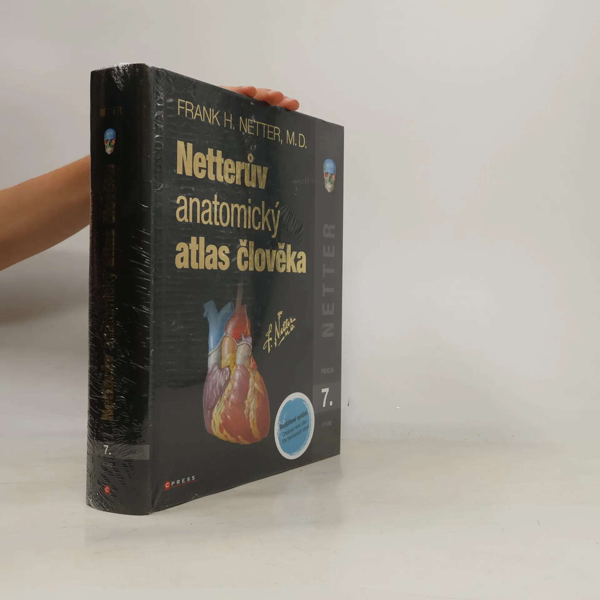
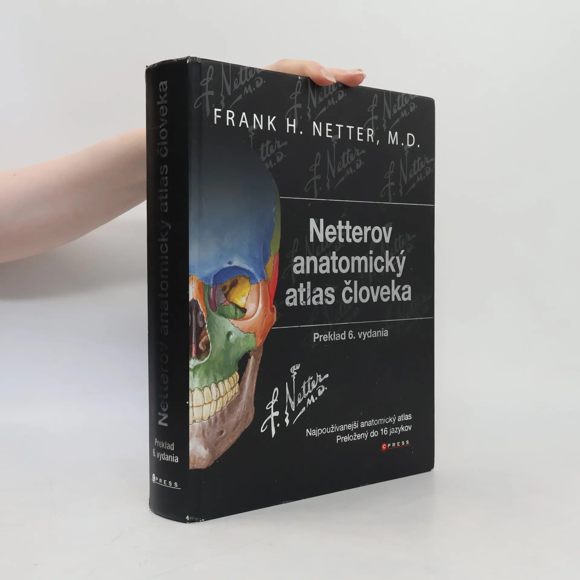
Netterův anatomický atlas člověka: Překlad 8. vydání
- 824bladzijden
- 29 uur lezen
Pro studenty a zdravotnické pracovníky, kteří studují anatomii, účastní se pitev či si osvěžují své znalosti, je tento anatomický atlas nenahraditelnou pomůckou. Představuje lidské tělo oblast po oblasti, v jasných a brilantních detailech, z pohledu klinického lékaře. Mezi ostatními je jedinečný, protože obsahuje ilustrace zdůrazňující anatomické struktury, jež jsou pro lékaře během studia i v praxi nejdůležitější. Zahrnuje více než 550 anatomických Tabulí a desítky pečlivě vybraných radiologických snímků.
Tento anatomický atlas človeka je vo svete uznávaný a považovaný za vrchol anatomickej vedeckej ilustrácie. Stal sa najpredávanejším anatomickým atlasom, ktorý je preložený už do 16 jazykov. Toto vydanie je prekladom 4. anglického vydania. Genialita obrázkov dr. Nettera spočíva v tom, že anatómiu zobrazuje jasne, realisticky, vedeckým spôsobom, a pritom zachováva vyváženosť medzi komplikovanosťou a prehnanou zjednodušenosťou. Toto vydanie obsahuje vyše 540 farebných a veľmi názorných ilustrácií, najnovšie informácie, rádiogramy, snímky počítačovej tomografie (CT), CT angiogramy a snímky magnetickej rezonancie. Anatomická terminológia bola v tomto vydaní doplnená tiež o najnovšie poznatky.
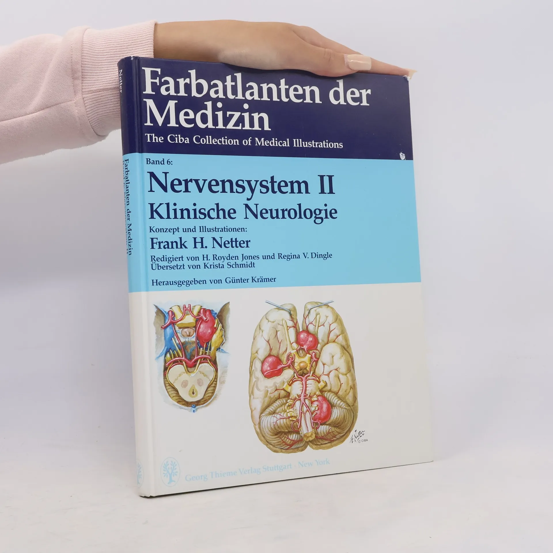
Nervensystem II. Klinische Neurologie
Farbatlanten der Medizin. Band 6
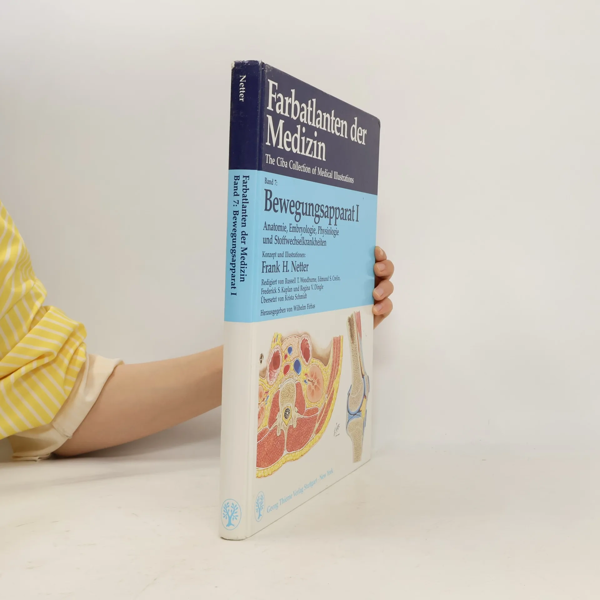
Farbatlanten der Medizin Band 7. Bewegungsapparat I.
Anatomie, Embryologie, Physiologie und Stoffwechselkrankheiten
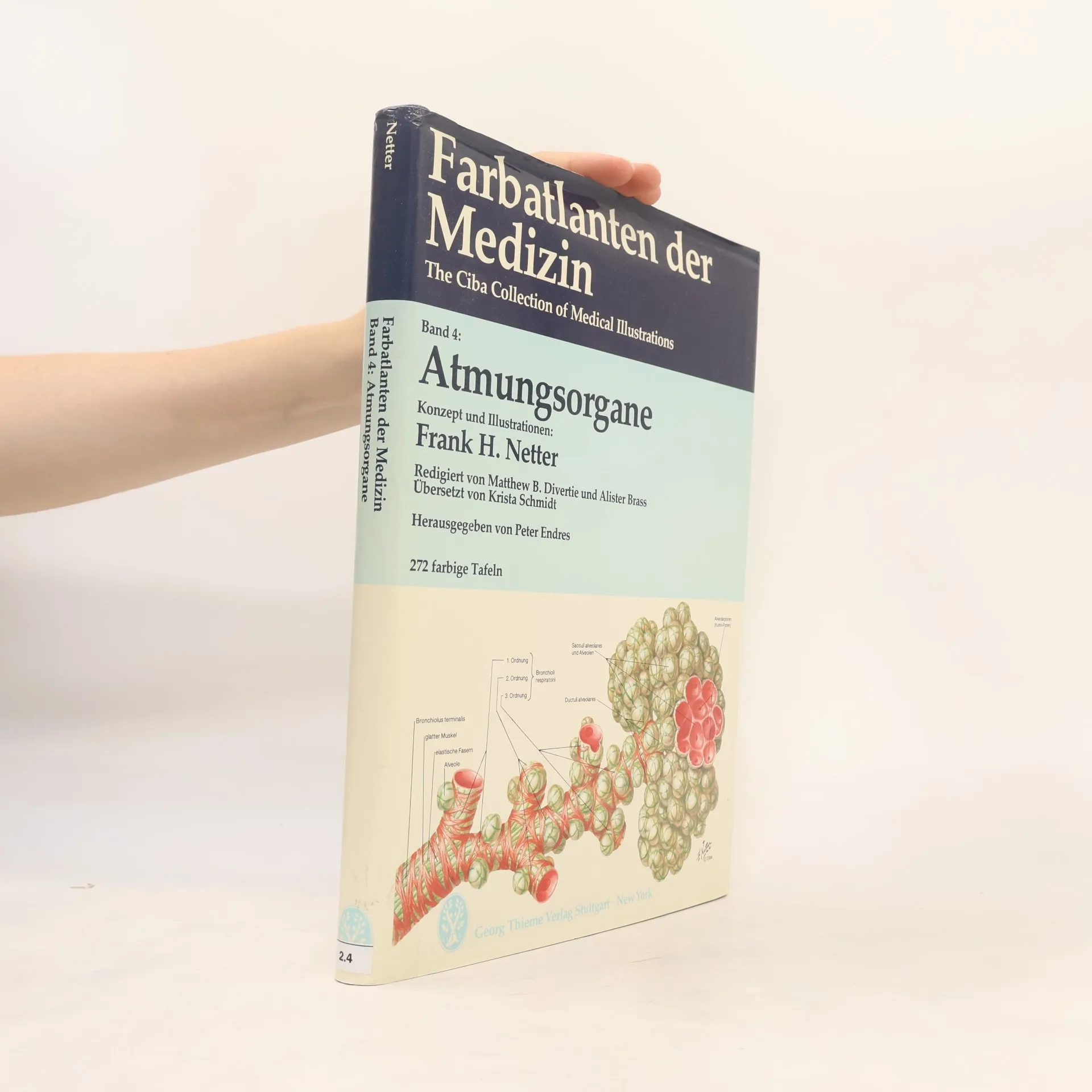
Farbatlanten der Medizin. Band. 4, Atmungsorgane
- 320bladzijden
- 12 uur lezen
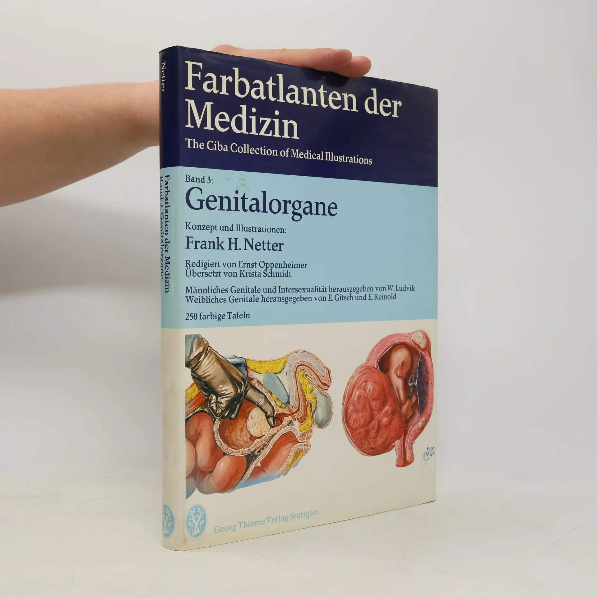
Farbatlanten der Medizin. Band 3: Genitalorgane
- 280bladzijden
- 10 uur lezen
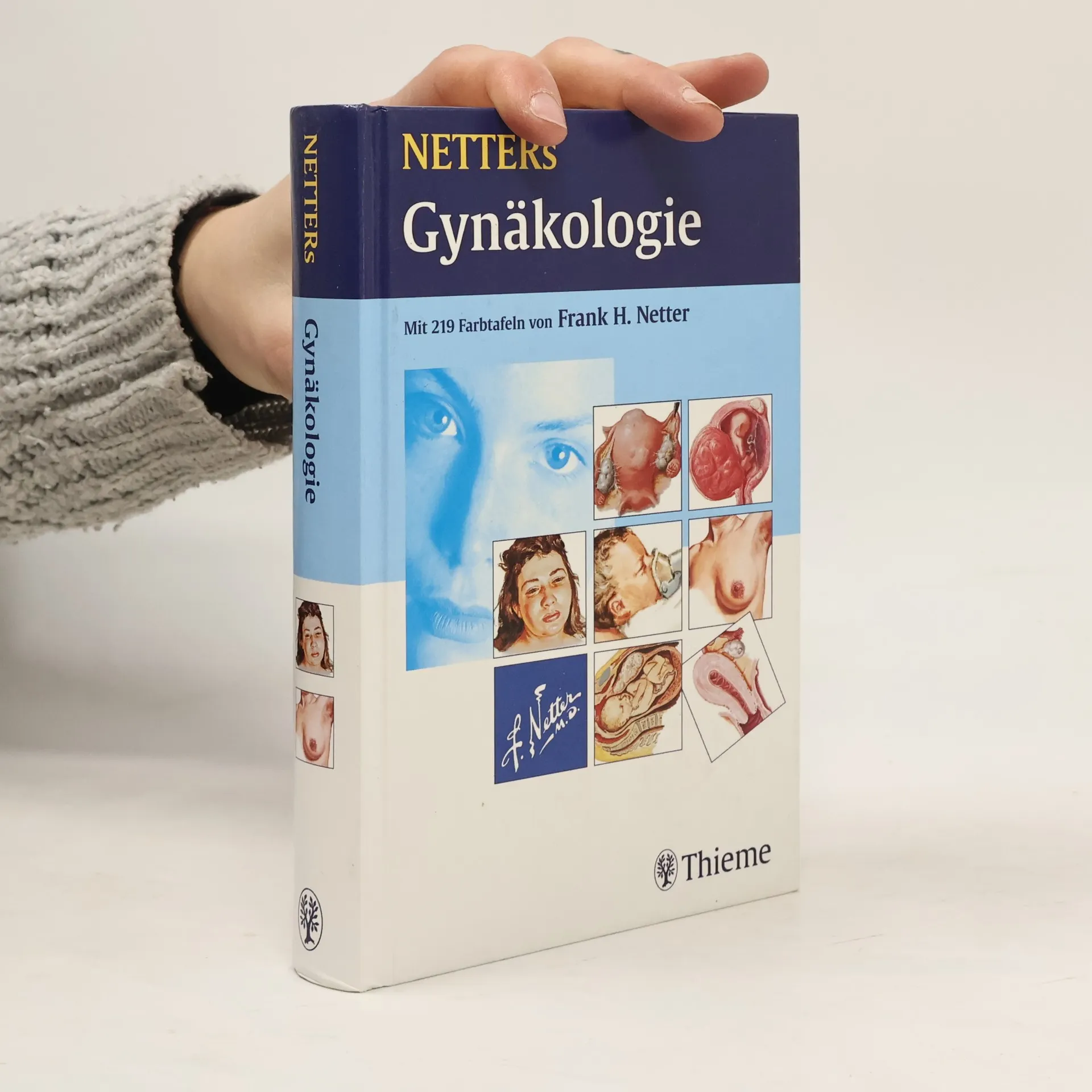
Netters Gynäkologie
- 473bladzijden
- 17 uur lezen
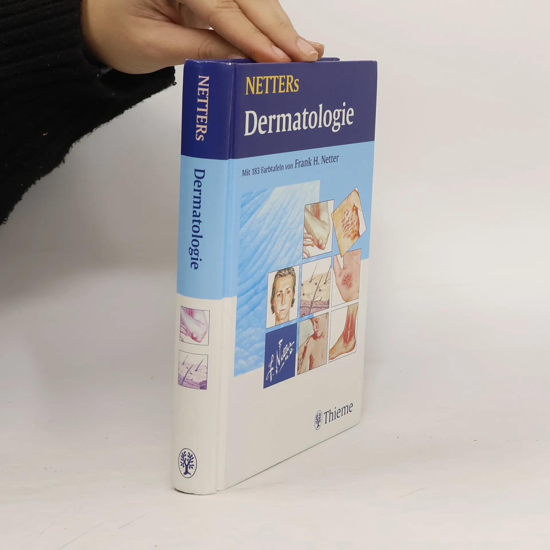
Netters Dermatologie
- 408bladzijden
- 15 uur lezen
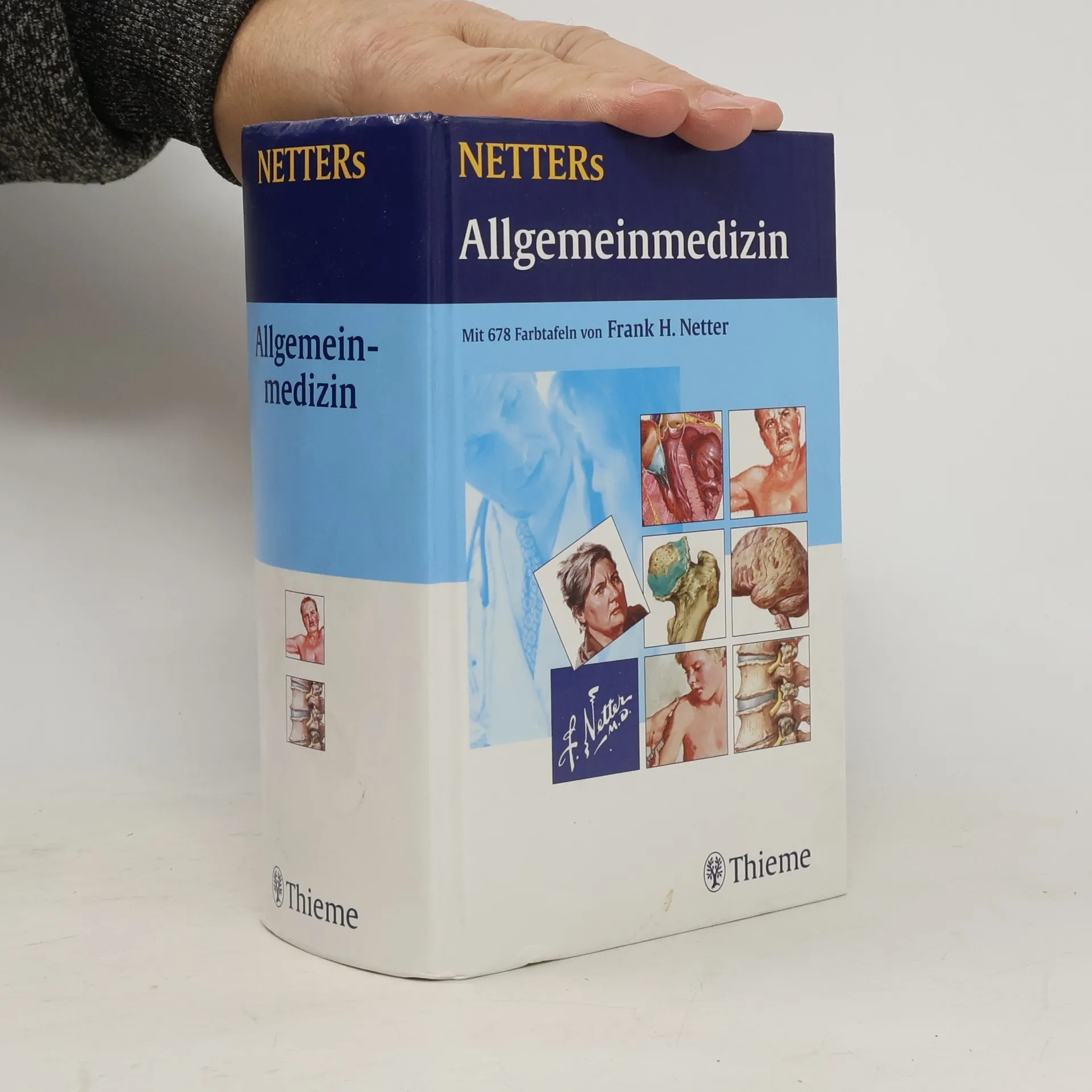
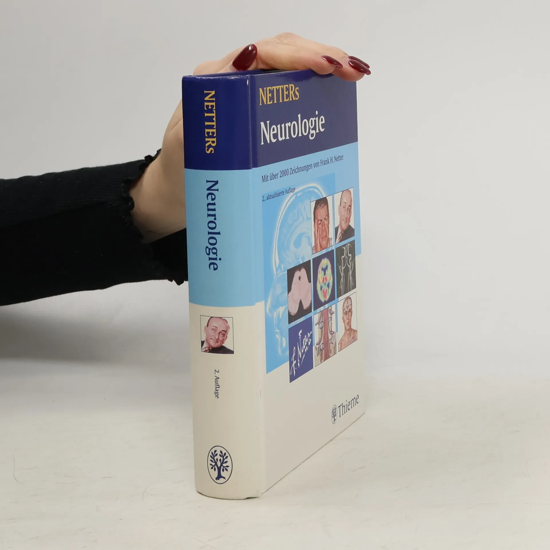
Netters Neurologie
- 535bladzijden
- 19 uur lezen
Netters Neurologie - der Klassiker jetzt noch besser Das gesamte Bild der Medizin - Einmaliger Überblick über alle neurologischen Krankheitsbilder - Mehr als 2000 Einzeldarstellungen auf über 500 Seiten - Der geniale Abbildungsschatz von Frank Netter im Taschenatlasformat - Knappe Erläuterungen und einprägsame Illustrationen veranschaulichen jedes Krankheitsbild - Anatomie, Physiologie und Klinik zusammen auf einer Bildtafel Alle Vorteile des visuellen Lernens - Zusammenhänge verstehen - ungewöhnliche Einblicke und einprägsame Kurztexte - Ganzheitliches Verständnis - Anatomie, Physiologie und Klinik zusammen auf einer Bildtafel - Bilder, die haften bleiben - ideale Ergänzung zum Lehrbuch Der Standard der Medizin - Präzise, einprägsam, didaktisch vollendet - Netters Bilder sind in Weiterbildung und Lehre seit Jahrzehnten die Nummer eins - weltweit - Die Vorlesung im Taschenbuch - ganze Ärztegenerationen sind mit diesen Bildern aufgewachsen Prof. O. Schneider: „Der Ärzteschaft ist Dr. Netter weltweit als Schöpfer auf das Wesentliche zielender, an der Klinik orientierter medizinischer Illustrationen von unendlicher Klarheit ein Begriff geworden.“
Netters Pädiatrie
- 591bladzijden
- 21 uur lezen
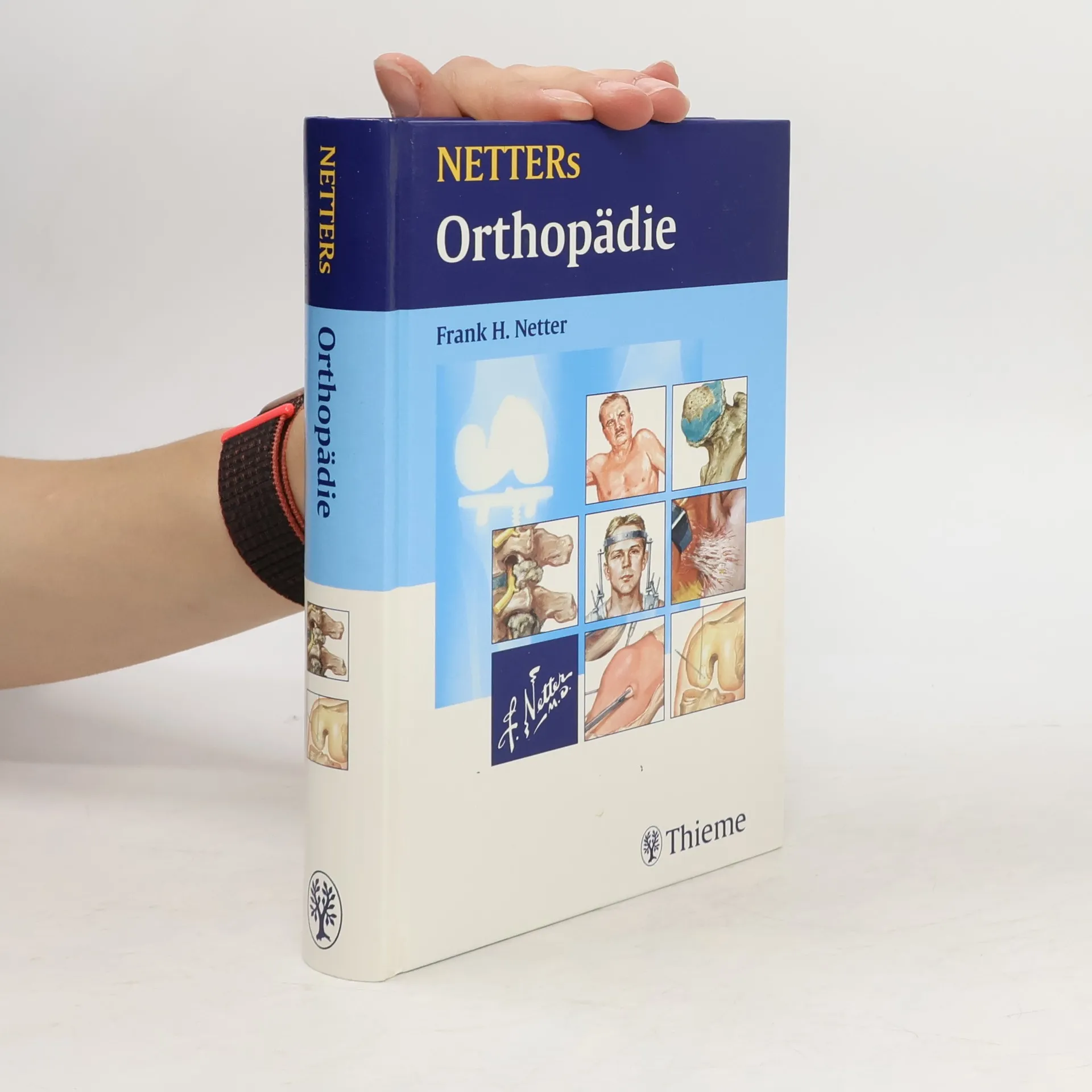
Netters Orthopädie
- 594bladzijden
- 21 uur lezen
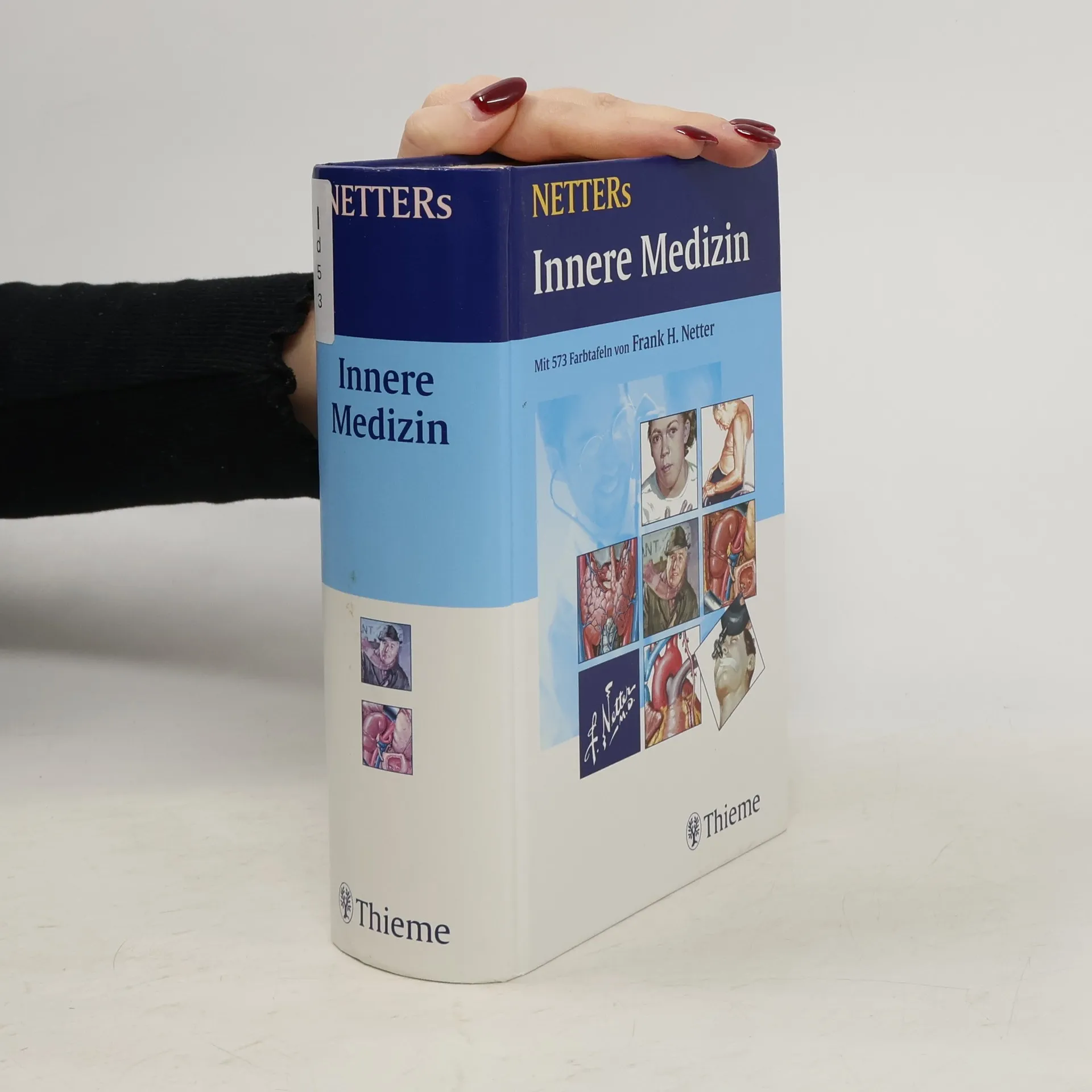
Netter's Innere Medizin
- 1175bladzijden
- 42 uur lezen
Umfassend! Über 1800 Abbildungen bieten einen einmaligen Überblick über alle internistischen Krankheitsbilder Anschaulich! Einprägsame Illustrationen und knappe Erläuterungen erklären jedes Krankheitsbild: verstehen, lernen und für immer einprägen Der schnelle Überblick! Anatomische und physiologische Grundlagen, Pathologie und Klinik, auf einer Bildtafel Aktuell: Einzigartige Netter-Bilder ergänzt um aktuelle bildgebende Diagnostik und aktuelle Therapie Perfekt! Besser können medizinische Illustrationen nicht sein: Didaktisch vollendet, präzise, einprägsam Das ist neu Komplette Überarbeitung von Diagnostik und Therapie Einzigartige Netter-Illustrationen ergänzt mit klinischen Befunden und aktueller Bildgebung Noch übersichtlicher: neues größeres Format
Das am meisten kritisierte Werk von Dr. Frank H. Netter, dieses vollständig illustrierte Buch aus der Referenzsammlung umfasst: Hunderte von weltweit renommierten Illustrationen von Frank H. Netter, MD; informative Texte von anerkannten medizinischen Experten; Anatomie, Physiologie und Pathologie; sowie diagnostische und chirurgische Verfahren.
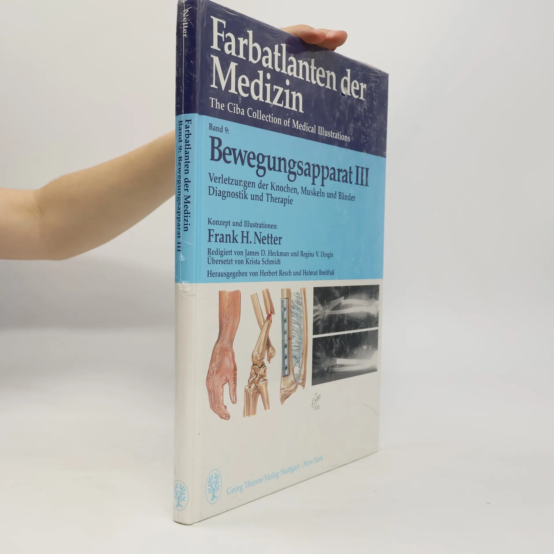
Farbatlanten der Medizin - 9: Bewegungsapparat
Verletzungen der Knochen, Muskeln und Bänder Diagnostik und Therapie
- 202bladzijden
- 8 uur lezen
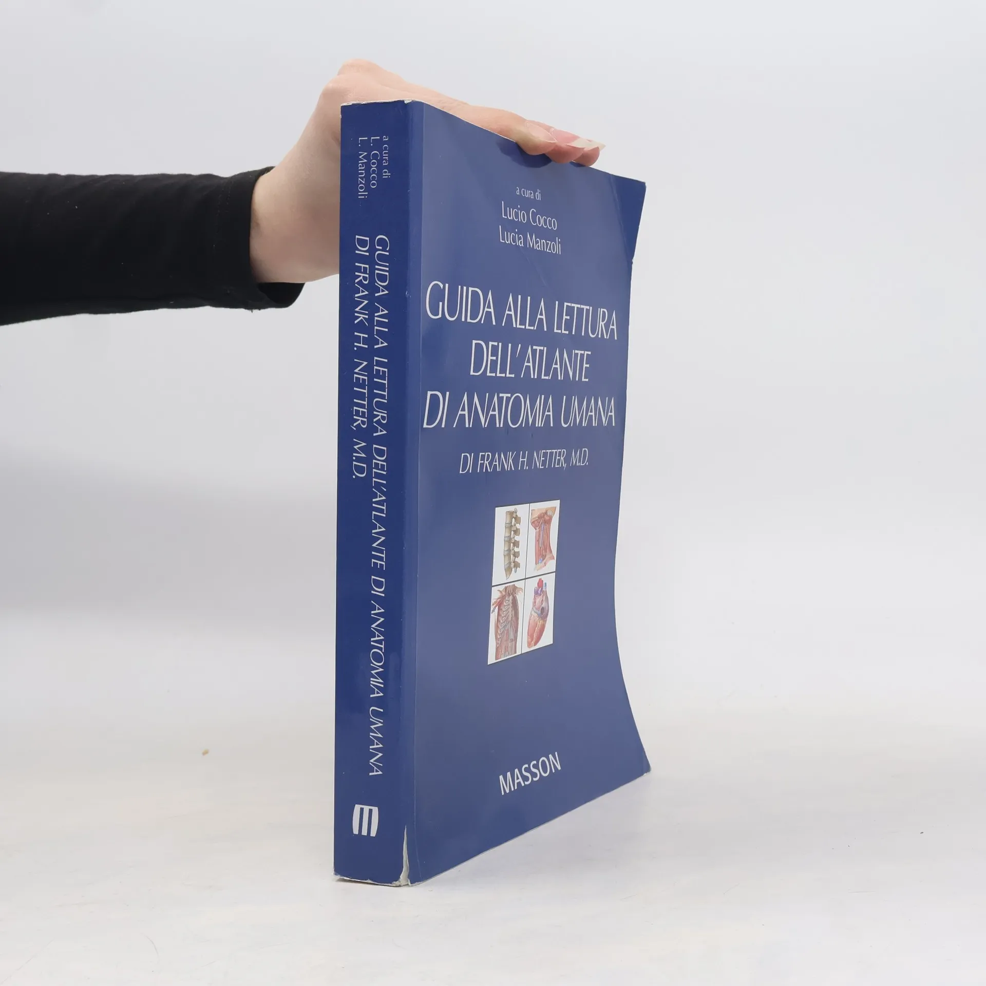
Guida alla lettura dell'atlante di anatomia umana di Frank H. Netter, M. D.
- 542bladzijden
- 19 uur lezen
Tegnede billedkort af menneskets anatomi med tekst på bagsiden, således at kortene kan bruges som vendekort i indlæringssituationer